Anatomy of the Human Shoulder Joint Biology Diagrams
Anatomy of the Human Shoulder Joint Biology Diagrams The main movement around the shoulder joint is to rotate the arm in a circular motion or to abduct out and away from the body, which is mainly assisted by the glenohumeral joint—also known as shoulder joint. Below we will reveal the shoulder joint anatomy. 1. Bones. The humerus and scapula are two main bones forming the shoulder joint anatomy.
:max_bytes(150000):strip_icc()/499158609-56a6d9935f9b58b7d0e51b84.jpg)
Learn about the anatomy of the shoulder joint, a ball and socket synovial joint that allows a wide range of movement. Find out how the joint is stabilised by ligaments, muscles, bursae and the glenoid labrum, and what are the common injuries and clinical relevance of the shoulder joint.
Anatomy of the Human Shoulder Joint Biology Diagrams
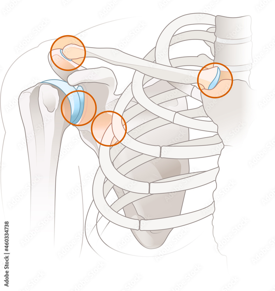
Learn about the shoulder joint complex, which consists of four articulations: glenohumeral, acromioclavicular, sternoclavicular and scapulothoracic. Find out the articular surfaces, ligaments, bursae, relations, arteries, nerves and movements of the shoulder joint.
Learn about the bones, muscles, ligaments, and movements of the shoulder joint, one of the most complex and mobile joints of the body. Find out how the shoulder joint can be affected by injuries, diseases, and conditions such as rotator cuff tear, frozen shoulder, and labral tear. Learn about the two joints that form the shoulder, the glenohumeral and acromioclavicular joints, and how they enable the arm to move in various directions. See 3D images and diagrams of the shoulder bones, muscles, ligaments, and cartilage.

Shoulder Joint Anatomy Biology Diagrams
Learn about the structure and function of the shoulder, the most mobile joint in the human body. The shoulder consists of three bones, four joints, ligaments, muscles, and nerves that enable a wide range of motion.
