Pelvic floor muscles Biology Diagrams
Pelvic floor muscles Biology Diagrams Learn about the muscles that form the lower limit of the true pelvis and support the pelvic organs. Find out their attachments, blood supply, innervation, function, embryology, and clinical significance. INTRODUCTION. Pelvic floor muscles have two major functions; they provide 1; support or act as a " floor" for the abdominal viscera including the rectum and 2; constrictor or continence mechanism to the urethral, anal and vaginal orifices (in females).Here, we will discuss the relevance of pelvic floor to the anal opening and closure function, and discuss new findings with regards to the
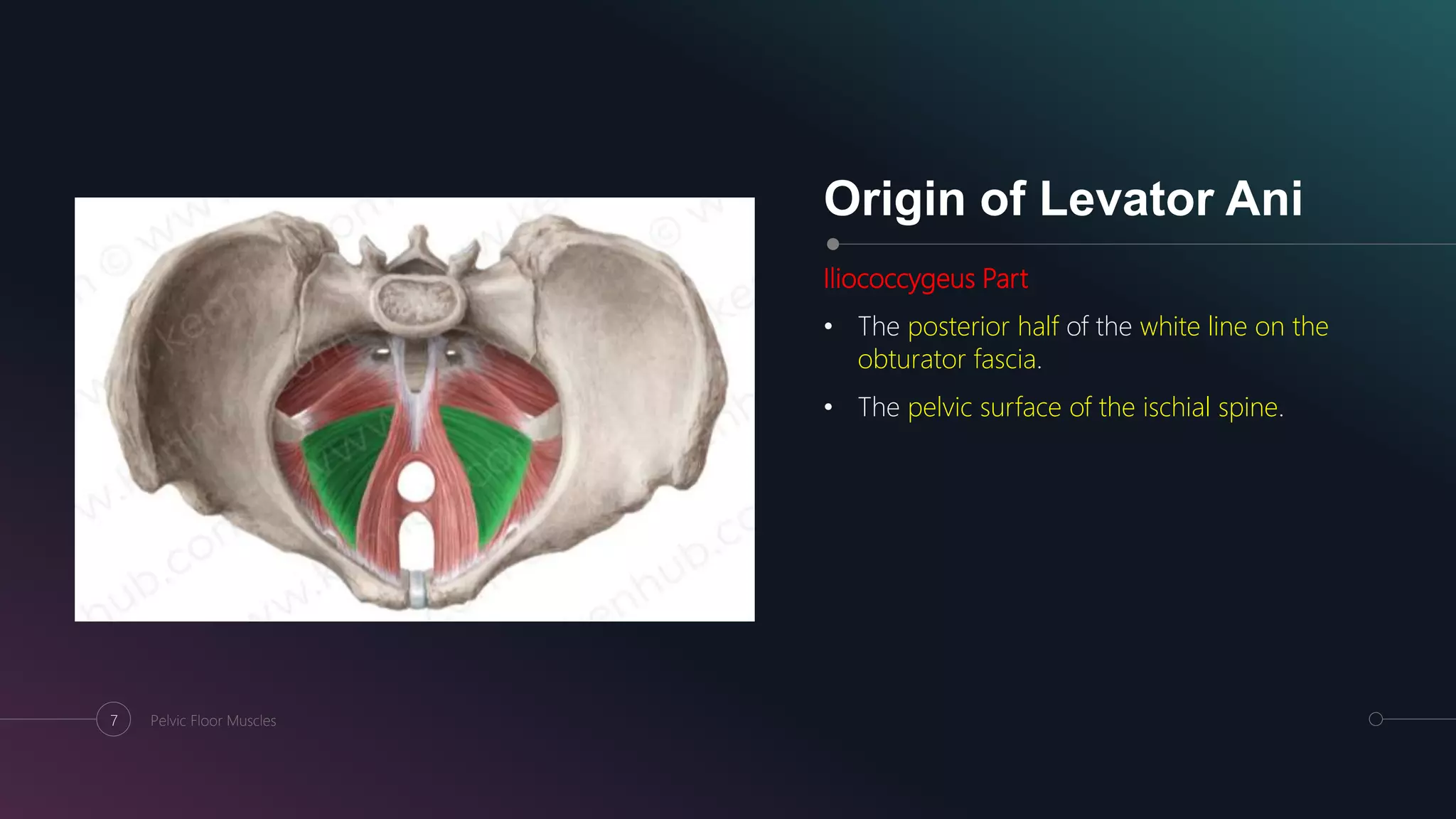
Learn about the anatomy and functions of the pelvic floor, a funnel-shaped structure that supports the pelvic viscera and maintains urinary and faecal continence. The pelvic floor consists of three main components: levator ani, coccygeus and fascia coverings.

Pelvic Floor Anatomy Biology Diagrams
Pelvic floor muscle training versus no treatment, or inactive control treatments, for urinary incontinence in women: a cochrane systematic review abridged republication. European Journal of Physical and Rehabilitation Medicine, 54(3), 416-432. ↑ Sapsford, R. (2001). Rehabilitation of pelvic floor muscles utilizing trunk stabilization.

Figure 43 illustrates the muscles of the pelvic floor. Pelvic floor work is often performed intrarectally or intravaginally and is therefore usually beyond the scope of practice for most manual therapists. However, some pelvic floor musculature (coccygeus and levator ani) are partially accessible from the outside, inferior to the piriformis
Muscles of the pelvic floor: Anatomy and function Biology Diagrams
Pelvic floor dysfunction refers to a set of indications and symptoms caused by faulty pelvic floor muscle activity. The pelvic floor muscles in women support the urethra, vagina, and anal canal. Weakness of these muscles can lead to a lack of structural support for these organs, manifesting as: